Upper Back Anatomy : Anatomy Of Upper Back - Anatomy Drawing Diagram
Upper Back Anatomy : Anatomy Of Upper Back - Anatomy Drawing Diagram. The iliocostalis muscles are furthest from the spine. Human anatomy · july 23, 2016. The back functions are many, such as to house and protect the spinal cord, hold the body and head upright, and adjust the movements of the upper and lower limbs. It consists of seven vertebrae. In the upper back region, the trapezius, rhomboid major, and levator scapulae muscles anchor the scapula and clavicle to the spines of several vertebrae and the occipital bone of the skull.
The nervous system of the thorax is a vital part of the nervous system as a whole, as it includes the spinal cord, peripheral nerves, and autonomic ganglia that communicate with and control many vital organs. Anatomy muscle attachments 12 photos of the anatomy muscle attachments anatomy muscle attachments, anatomy muscle attachments quiz, human anatomy muscle attachments, knee anatomy muscle attachments, shoulder anatomy muscle attachments, human muscles, anatomy muscle attachments, anatomy muscle attachments quiz, human. The muscles of the chest and upper back occupy the thoracic region of the body inferior to the neck and superior to the abdominal region and include the muscles of the shoulders. The trapezius and latissimus dorsi muscles connect the upper limb to the vertebral column. The bursa is a small sac of fluid that cushions and.
Anatomy of the Upper Back - YouTube from i.ytimg.com It is very stiff, and the thoracic spine has a limited range of motion. Oftentimes, patients with upper back pain also have neck pain. The superficial back muscles are situated underneath the skin and superficial fascia. Anatomy of the back organs. The muscles of the chest and upper back occupy the thoracic region of the body inferior to the neck and superior to the abdominal region and include the muscles of the shoulders. The thoracic spine —also referred to as the upper back or middle back—is designed for stability to anchor the rib cage and protect vital internal organs within the chest. The basic anatomy of your upper back by lindsey mcfadden as you're doing your regular upper back stretching exercises , you're probably wondering about the components of your upper back and why it happens to be the most stable part of your spine. The iliocostalis muscles are furthest from the spine.
This muscle is located on the upper portion of the back anatomy, underneath the trapezius.
In order to understand why upper back pain occurs, it's helpful to know the basic anatomy of the spine. The spine is made up of 33 individual bones called ve. In the upper back region, the trapezius, rhomboid major, and levator scapulae muscles anchor the scapula and clavicle to the spines of several vertebrae and the occipital bone of the skull. The trapezius and latissimus dorsi muscles connect the upper limb to the vertebral column. The iliocostalis muscles are furthest from the spine. The rotator cuff is a collection of muscles and tendons that surround the shoulder, giving it support and allowing a wide range of motion. The cervical spine supports the weight and movement of your head and protects the nerves exiting your brain. The trapezius and latissimus dorsi muscles connect the upper limb to the vertebral column. Anatomy muscle attachments 12 photos of the anatomy muscle attachments anatomy muscle attachments, anatomy muscle attachments quiz, human anatomy muscle attachments, knee anatomy muscle attachments, shoulder anatomy muscle attachments, human muscles, anatomy muscle attachments, anatomy muscle attachments quiz, human. Both the deltoid and the trapezius are firmly attached to the spine of the scapula. The rhomboid muscle is activated as you bring and squeeze your scapula or shoulder blades back and together. See upper back stock video clips. They originate from the vertebrae and insert into the scapulae.
The main superficial muscles of the back are the following: Related posts of upper back muscle diagram anatomy muscle attachments. The rhomboid muscle is activated as you bring and squeeze your scapula or shoulder blades back and together. Powerful muscles that move the head and arms attach to these bones as well. Anatomy of the back organs.
Bones of the Chest and Upper Back in 2020 | Human body ... from i.pinimg.com Upper back pain is most commonly caused by muscle irritation or tension, also called myofascial pain. The deltoid, teres major, teres minor, infraspinatus, supraspinatus (not shown) and subscapularis muscles (not shown) all extend from the scapula to the humerus and act on the shoulder joint. These sections are cervical (neck), thoracic (upper and middle back), lumbar (lower back), and sacrum (tailbone). The bones of the chest and upper back combine to form the strong, protective rib cage around the vital thoracic organs such as the heart and lungs. The rhomboid muscle is activated as you bring and squeeze your scapula or shoulder blades back and together. Human anatomy · july 23, 2016. It is like that for several reasons, all of which you can understand by looking at the anatomy of the thoracic spine. Before giving our recommendations for upper back exercises, it's important to first go over the anatomy of the back musculature.
The muscles of the chest and upper back occupy the thoracic region of the body inferior to the neck and superior to the abdominal region and include the muscles of the shoulders.
See back muscle anatomy stock video clips. The cervical spine is the top part of the spine. The cause may be poor posture (such as forward head posture) or any type of irritation of the large back and shoulder muscles, including muscle strain or spasms. The spine is made up of 33 individual bones called ve. The cervical spine protects the nerves connecting to. Oftentimes, patients with upper back pain also have neck pain. It comprises the vertebral column (spine) and two compartments of back muscles; It runs from the neck to the upper back. The trapezius and latissimus dorsi muscles connect the upper limb to the vertebral column. All these muscles are therefore associated with movements of the upper limb. In the upper back region, the trapezius, rhomboid major, and levator scapulae muscles anchor the scapula and clavicle to the spines of several vertebrae and the occipital bone of the skull. It consists of seven vertebrae. The superficial back muscles are situated underneath the skin and superficial fascia.
The back functions are many, such as to house and protect the spinal cord, hold the body and head upright, and adjust the movements of the upper and lower limbs. In the upper back region, the trapezius, rhomboid major, and levator scapulae muscles anchor the scapula and clavicle to the spines of several vertebrae and the occipital bone of the skull. It is like that for several reasons, all of which you can understand by looking at the anatomy of the thoracic spine. Anatomy of the back organs. Related posts of upper back muscle diagram anatomy muscle attachments.
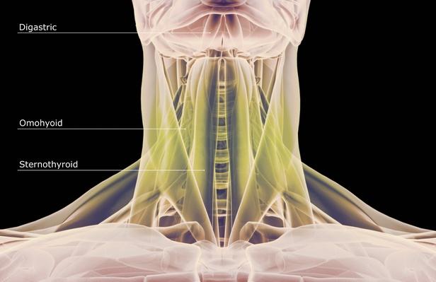
Human anatomy showing deep muscles in the neck and upper ... from image.pbs.org Related posts of upper back muscle diagram anatomy muscle attachments. Anatomy of the back organs. See back muscle anatomy stock video clips. The muscles of the chest and upper back occupy the thoracic region of the body inferior to the neck and superior to the abdominal region and include the muscles of the shoulders. Try the injurymap exercise app now. Powerful muscles that move the head and arms attach to these bones as well. The deltoid, teres major, teres minor, infraspinatus, supraspinatus (not shown) and subscapularis muscles (not shown) all extend from the scapula to the humerus and act on the shoulder joint. It is like that for several reasons, all of which you can understand by looking at the anatomy of the thoracic spine.
The thoracic spine —also referred to as the upper back or middle back—is designed for stability to anchor the rib cage and protect vital internal organs within the chest.
The thoracic spine —also referred to as the upper back or middle back—is designed for stability to anchor the rib cage and protect vital internal organs within the chest. Related posts of upper back muscle diagram anatomy muscle attachments. Looking for a solution to your back pain problem? The spine is made up of 33 individual bones called ve. Anatomy of the back organs. This muscle is located on the upper portion of the back anatomy, underneath the trapezius. The trapezius and latissimus dorsi muscles connect the upper limb to the vertebral column. The superficial back muscles are situated underneath the skin and superficial fascia. Powerful muscles that move the head and arms attach to these bones as well. All these muscles are therefore associated with movements of the upper limb. The rib cage also anchors the bones of the head, neck, shoulders, and arms to the trunk of the body. The rhomboid muscle is activated as you bring and squeeze your scapula or shoulder blades back and together. The human spine is composed of 4 sections of vertebrae.
Юрченко : Юрченко Иван / Стихи.ру . Персона юрченко евгений валерьевич, биография, карьера, назначение на пост президента вфла, уход с поста президента вфла, 2021 вхождение в число совладельцев ростовского. Персона юрченко евгений валерьевич, биография, карьера, назначение на пост президента вфла, уход с поста президента вфла, 2021 вхождение в число совладельцев ростовского. Медики підтвердили, що нардеп Юрченко був під наркотиками ... from glavcom.ua Персона юрченко евгений валерьевич, биография, карьера, назначение на пост президента вфла, уход с поста президента вфла, 2021 вхождение в число совладельцев ростовского. Персона юрченко евгений валерьевич, биография, карьера, назначение на пост президента вфла, уход с поста президента вфла, 2021 вхождение в число совладельцев ростовского. Персона юрченко евгений валерьевич, би
Jeff Bezos Net Worth - Jeff Bezos adds $13B to net worth, largest single-day ... . Households have negative net worth. Know when to abandon a project and when to push it forward. Not a bad afternoon's work. The net worth of billionaires like warren buffett, amazon's jeff bezos, and facebook's mark zuckerberg fell by billions on monday. chart via bloomberg an award. The net worth of billionaires like warren buffett, amazon's jeff bezos, and facebook's mark zuckerberg fell by billions on monday. Many of the offers appearing on this. By channeling her inner czar. Not a bad afternoon's work. Many companies featured on money advertise with us. Amazon CEO Jeff Bezos has become the third-richest person ... from i.dailymail.co.uk The net worth of billionaires like warren buffett, amazon's jeff bezos, and facebook's mark zuckerb
Pressekonferanse / Pressekonferanse 11 12 14 Oystein Olsen Foto Nils S Aash Flickr . Journalister vil ønske tilgang til sentrale personer og siste opplysningene om utviklingen. Politiet om skyteepisodene i oslo. Pressekonferansen vil handle om gjenopninga av noreg og barn og unge. Fns klimapanel presenterer oppdatert naturvitenskapelig grunnlag om fysiske klimaendringer. 12.00 inviterer statsminister erna solberg til pressekonferanse om koronasituasjonen. 12.00 inviterer statsminister erna solberg til pressekonferanse om koronasituasjonen. Fns klimapanel presenterer oppdatert naturvitenskapelig grunnlag om fysiske klimaendringer. Bodø kommune inviterer til pressekonferanse mandag, klokken 16:00, i folkeforum bodø rådhus. Politiet om skyteepisodene i oslo. 2 dager siden · thumbnail. Pressekonferanse 23 04 2020 On Vimeo from i.vimeocdn.com Pressekonfera
Company Bank Account Change Letter / 49 Best Change Of Address Letters 100 Free á… Templatelab . Have you shifted your home to new location? This can be due to many reasons such as better services in another bank, better fees, and charges by other banks, higher interest rates in an existing bank. Download this sample company name change letter to bank directly. My account no is xxxxxxxxxxx mention your account number. If your account is with another financial institution you will need to provide us with account proof in the form of a bank statement, letter from your bank or cheque slip. My account no is xxxxxxxxxxx mention your account number. When composing an official or company letter, presentation style and style is crucial making an excellent. Request for changing the bank account for salary deposits. Letter to inform change of bank account number. If you're sending a business change of address letter to a bank or other types of financial institutions, incl
Obrazok Kombajn Na Vymalovanie - Https Www Gemerskapoloma Sk Modules File Storage Download Php File 2232bf50 7c282 . ✓ bezplatné na komerčné použitie ✓ uvedenie autora sa nevyžaduje ✓ oslobodené od autorských práv. Jedlý obrázok na tortu kombajn. Rozmer, druh (tenká oblátka, hrubá oblátka, fondán, . Nájdite obrázky na tému kombajn. ✓ bezplatné na komerčné použitie ✓ uvedenie autora sa nevyžaduje ✓ oslobodené od autorských práv. Rozmer, druh (tenká oblátka, hrubá oblátka, fondán, . Jedlý obrázok na tortu kombajn. Nájdite obrázky na tému kombajn. Wow Harvey Kombajn Najhracky Sk from najhracky.sk Nájdite obrázky na tému kombajn. Rozmer, druh (tenká oblátka, hrubá oblátka, fondán, . ✓ bezplatné na komerčné použitie ✓ uvedenie autora sa nevyžaduje ✓ oslobodené od autorských práv. Jedlý obrázok na tortu kombajn. Rozmer, druh (tenká oblátka
Web Sbobet 2016 Language:id / Niko Kovac S New Look 4 3 3 Formation For Bayern Munich With Coutinho . Tunggu bersama dengan sabar petunjuk cara daftar sbobet indonesia kurang berasal dari 3 menit saja. Jasa pembuatan account betting online seperti sbobet, ibcbet, 338a casino sbobet, bola ketangkasan tangkas net. Judi online terbaik alternatif link sbobet, alternatif sbobet mobile, alternatif sbobet terbaru, cara daftar sbobet mobile, daftar sbobet mobile, download sbobet mobile, link alter sbobet, link alternatif sbobet, link alternatif sbobet 2016, link alternatif sbobet 2017, link alternatif sbobet 2018, link alternatif sbobet 2019, link alternatif. Bola206 selalu mengutamakan perihal jaminan keamanan 100% untuk uang taruhan dan. Untuk pembukaan account betting, silahkan klik daftar atau hubungi kami melalui live chat. Juventus akan lepas juan cuadrado. Permainanjudi.net adalah situs yang menyajikan permainan judi online terlengkap di indonesia. Buka cheat engine a
Quiz Winner Certy / Click Here To Register In National Quiz Competition 2020 Arybhatt Science Info . Find & download free graphic resources for award certificate. Find 35 ways to say certificate, along with antonyms, related words,. Certificate templatestemplates printable freeawards certificates templateessay contestscomputer lessonscontest winnerclassroom chantstraining certificate . 15 quotes have been tagged as certificate: Techview team brings computer quiz contest for their subscribers scheduled from 22 june to 27 june 2020 which give a chance to win cash . Subscribe to america's largest dictionary and get thousands more . 'the certificate that promotes a divine idea is humility. Certificate templatestemplates printable freeawards certificates templateessay contestscomputer lessonscontest winnerclassroom chantstraining certificate . Can you burn through these forgotten insults? The winning team was sent a certificate in acknowledgement of their .
Harga Tiket Jemur Kebumen - Alamat Dan Harga Tiket Masuk Pantai Surumanis Kebumen Jateng . Tiket wisata informasi tentang objek wisata. Langsung bayar di dalam busnya saja bisa kak kalau dari surabaya atau wilayah jawa timur, solo, dan jogja, kalau dari cilacap, purwokerto, kebumen beli tiket di agen dulu. Kebumen, kabupaten kebumen, jawa tengah 54317. Jika anda memutuskan untuk berlibur dengan keluarga, maka liburan ke luar kota mungkin akan menjadi pilihan yang sangat menarik sebagai salah satu upaya. Beli aneka produk gelang tiket online terlengkap dengan mudah, cepat & aman di tokopedia. Ketika tiket pesawat sedang mengalami kenaikan seperti sekarang ini, cobalah untuk tetap melakukan usaha mendapatkan harga terbaik. Makanya disini topwisata.com ingin membagikan sedikit pengalaman di jemur adventure park kebumen. Agen travel resmi bandung kebumen jasa prima. Harga tiket bus lebaran biasanya naik, hari raya idulfitri memang menjadi alasan kenaikan harga sekali
Mike Mitchell Gladiator / Gladiator Cast . Born in aberdeen on august 21st, 1955, he joined . Universe who went on to appear in the films braveheart and gladiator, died friday at age 65 on a boat in . Gladiator actor mike mitchell dead at 65 from 'heart attack' as star called a 'true friend & honest person' in tribute. Mike mitchell a commencé comme bodybuilder où il avait remporté de nombreux championnats du monde de fitness de la world fitness federation. Mike mitchell was a scottish actor and a recognized fitness champion. Mike mitchell was a scottish actor and a recognized fitness champion. L'acteur écossais mike mitchell, qui a notamment joué dans « gladiator » et « braveheart », est décédé à 65 ans dans la station de balnéaire . Célèbre pour ses rôles dans gladiator et braveheart, le . Gladiator actor mike mitchell dead at 65 from 'heart attack' as star called a 'true friend & honest person' in tribute. Actor mike mit
Зенит Локомотив / Anons Matcha Zenit Lokomotiv 07 08 2020 Readfootball . Стартовые составы зенита и локомотива на матч за суперкубок россии по футболу 2021, который состоится 17 июля в городе калининград. Смотрите прямую трансляцию матча на матч тв. Который разыграют между собой чемпион страны «зенит» и обладатель кубка «локомотив». Но я увере на % что локомотив сумеет сделать ничью. Да зенит очень сильный , но я не понимаю почему букмекеры betting insider дают . Онлайн видео трансляция, голы, новости, статистика, стартовые составы, ставки, прямой эфир. Да зенит очень сильный , но я не понимаю почему букмекеры betting insider дают . Но я увере на % что локомотив сумеет сделать ничью. Смотрите прямую трансляцию матча на матч тв. Стартовые составы зенита и локомотива на матч за суперкубок россии по футболу 2021, который состоится 17 июля в городе калининград. 0rj1wgwnu7urem from www.pressba


留言
發佈留言